
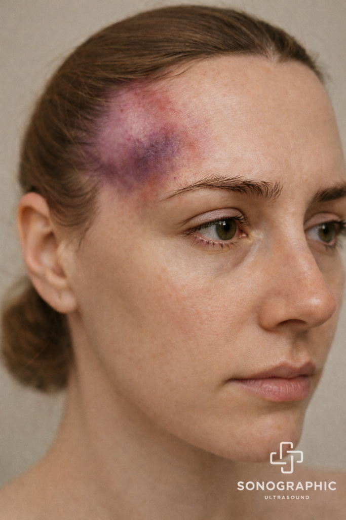
Using facial ultrasound during temple filler injections allows your provider to visualise blood vessels and tissue layers in real time. This imaging helps ensure the filler is placed safely and reduces the risk of vascular complications, which can affect the skin or, in very rare cases, vision.
A pre-treatment facial mapping ultrasound can be performed before your procedure. This step identifies the exact location of key blood vessels in your temples and helps your injector plan a safe injection path. Pre-treatment mapping is a proactive measure that significantly improves safety for your procedure.

To help keep you safe and well after your treatment, we provide a downloadable temple filler complications tracking card. This card makes it easy to record important details about your treatment, recognise early warning signs, and note any changes you experience after your procedure.
We recommend keeping this card with you following treatment and bringing it to any follow-up or urgent appointments, as it helps healthcare professionals quickly understand what treatment you have received and provide the right care if needed.
Designed to support clinical documentation, patient education, and risk management, the card assists practitioners in meeting professional and regulatory expectations for adverse event awareness, escalation, and record-keeping. It also empowers patients with clear guidance on what to monitor and when to seek clinical review, reinforcing a shared commitment to safe, accountable aesthetic care.
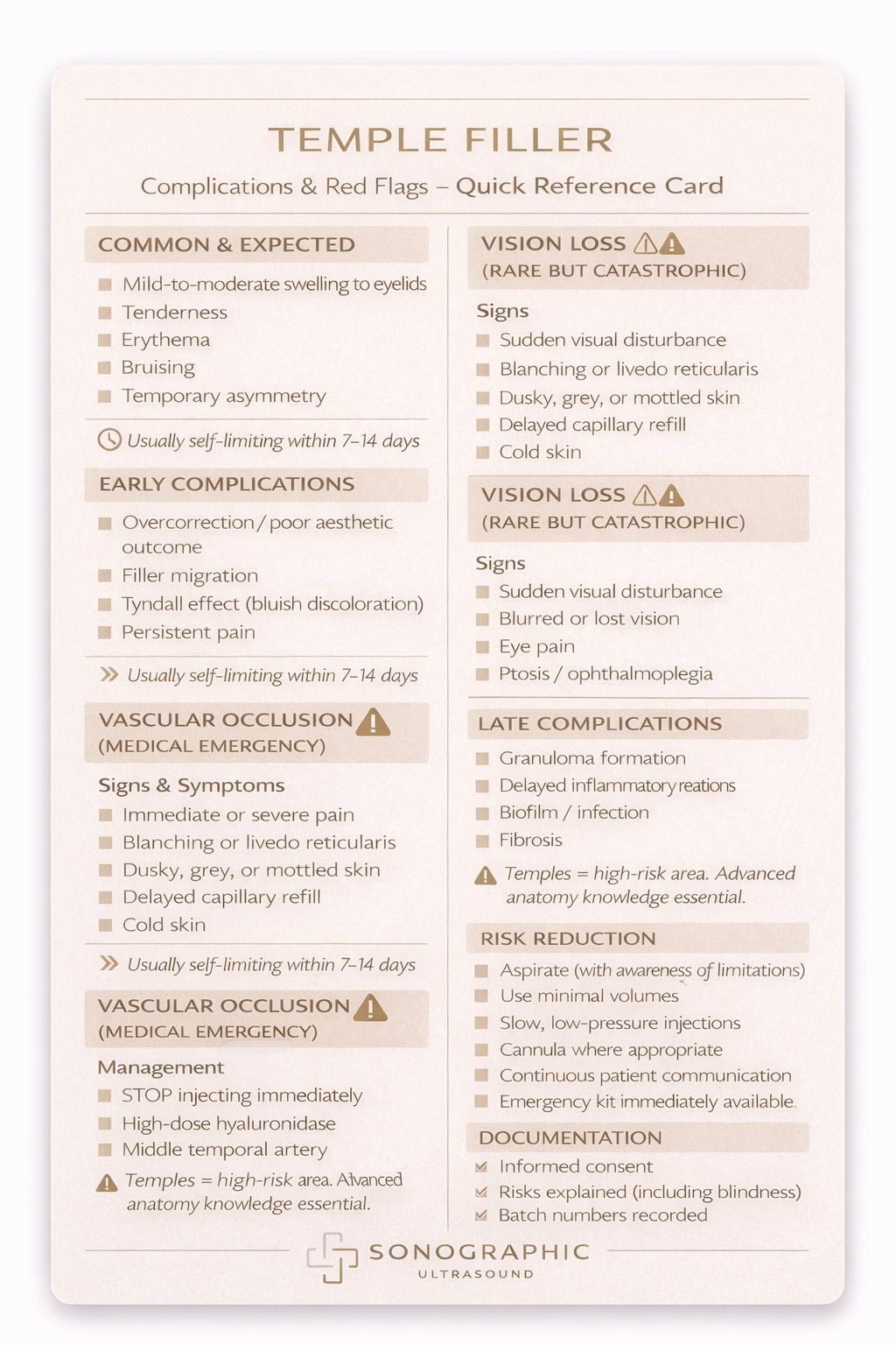
This quick reference guide is provided for educational and informational purposes only and is intended for use by appropriately trained medical professionals. It does not constitute medical advice, formal clinical guidance, or a substitute for accredited training, individual clinical judgement, or local regulatory protocols.
Aesthetic procedures carry inherent risks, including rare but serious complications such as vascular occlusion and vision loss. Practitioners must have appropriate training in facial anatomy, complication recognition, and emergency management, including immediate access to appropriate reversal agents and established referral pathways.
Sonographic Ultrasound accepts no liability for clinical decisions made based on this material. Clinicians are responsible for practising within their scope, professional guidelines, and indemnity requirements.
WhatsApp us